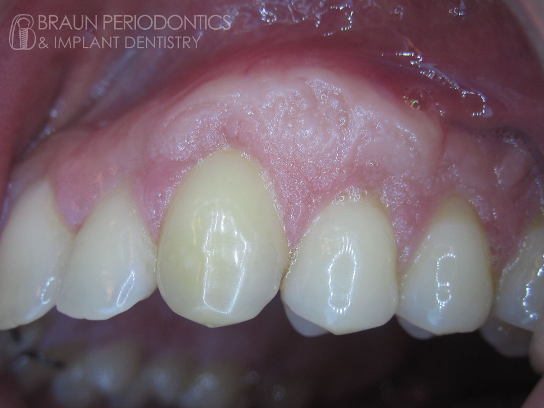
After: Roots covered with thick gum tissue
Before: Recessions on upper teeth
A photographic tour of some of our happy patients.
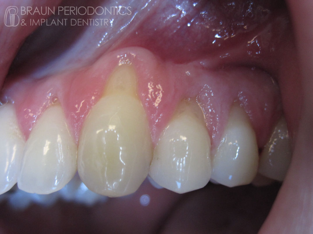
After: Roots covered with thick gum tissue
Before: Recessions on upper teeth
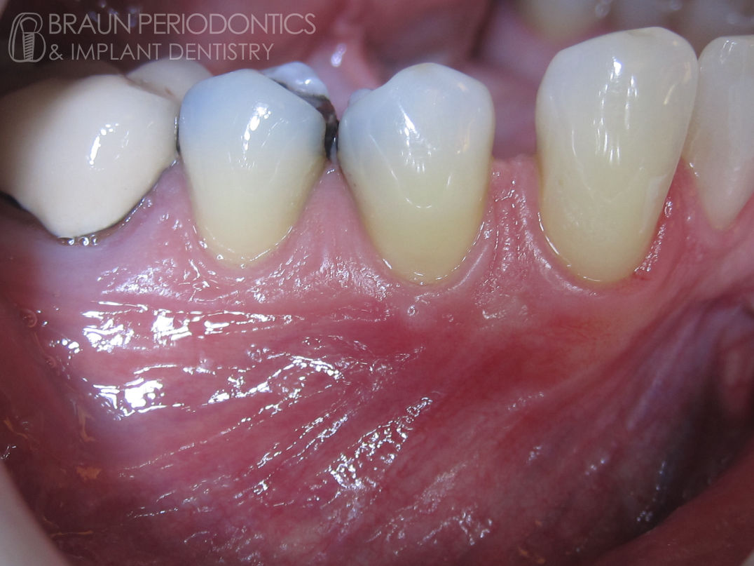
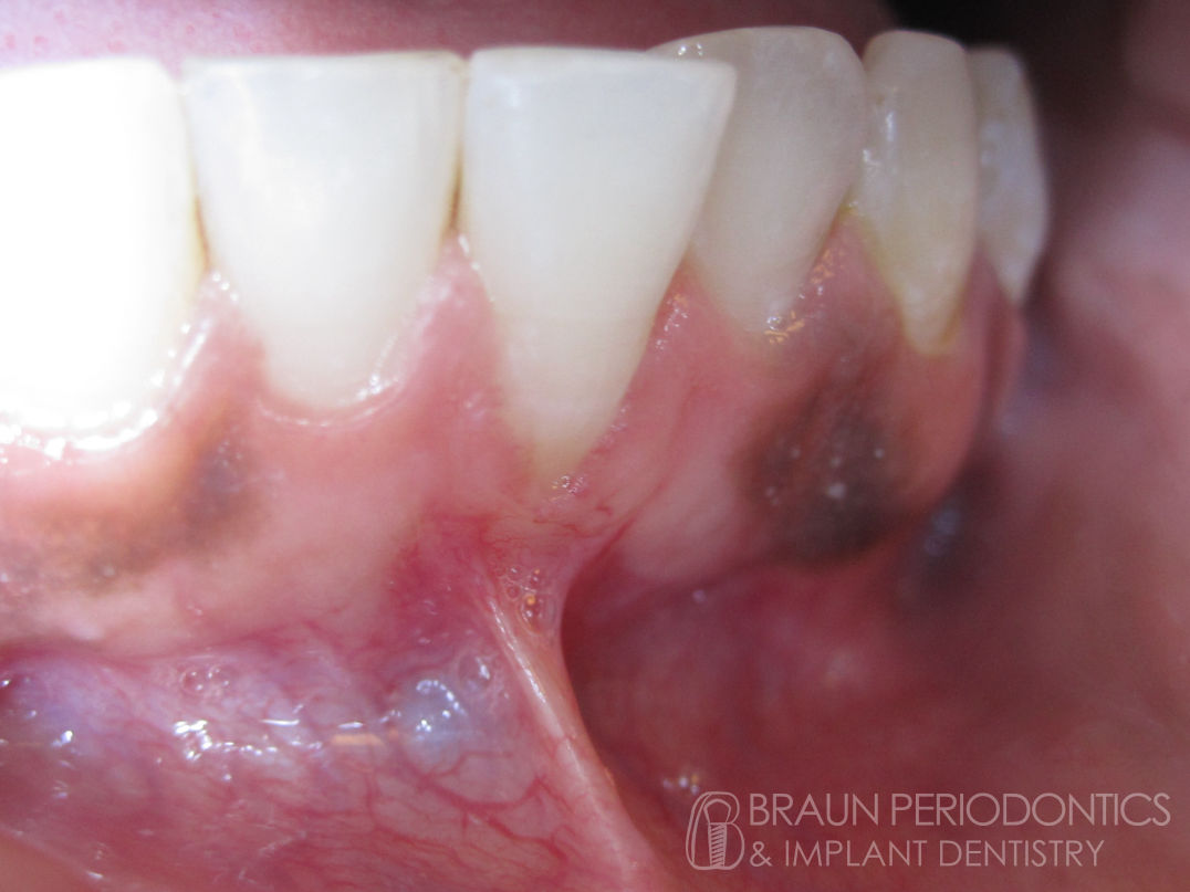
After: Frenum removed, grafted thick border of tissue
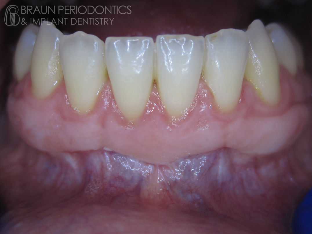
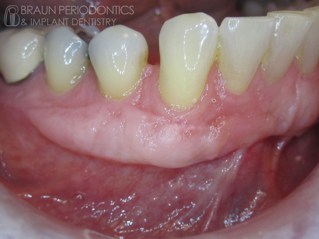
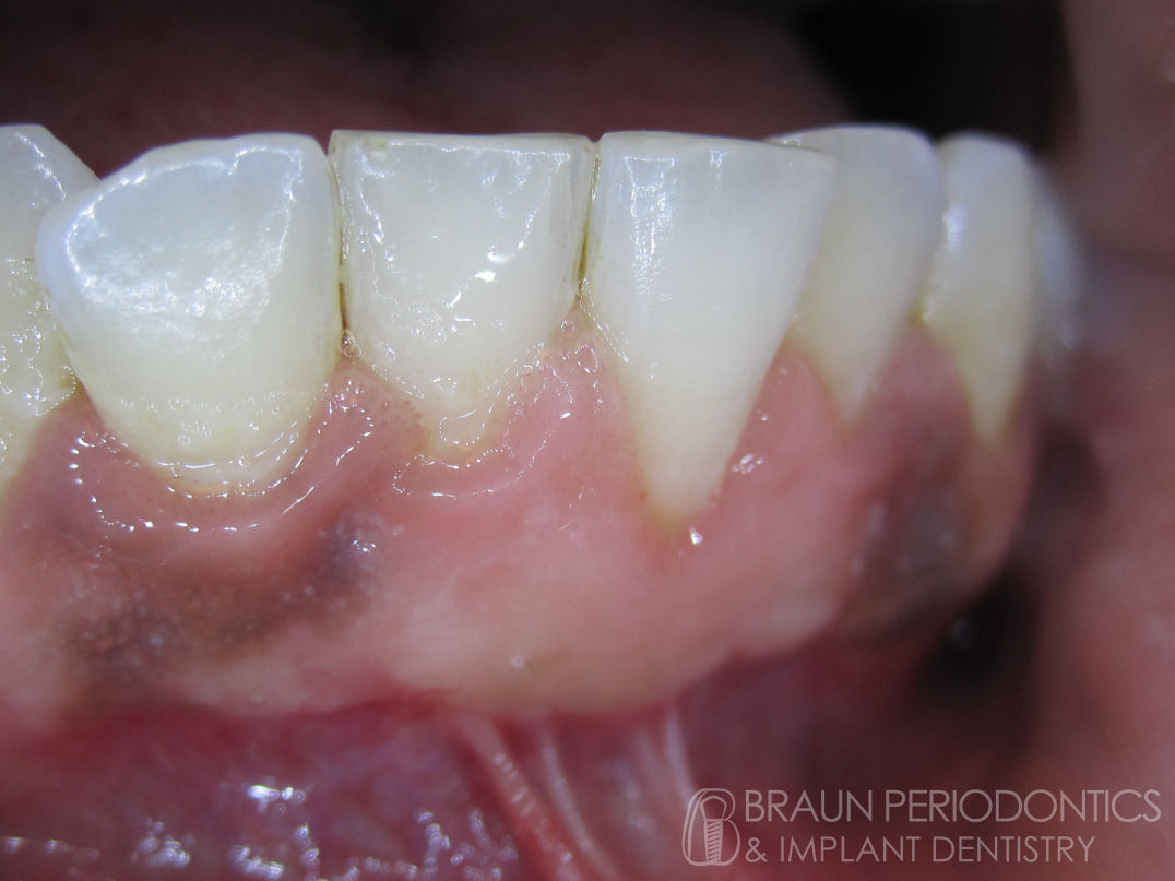
Before: Thin tissue and frenum along bottom teeth
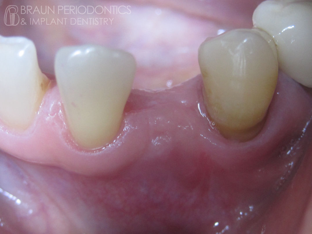
After: Thick strong border formed
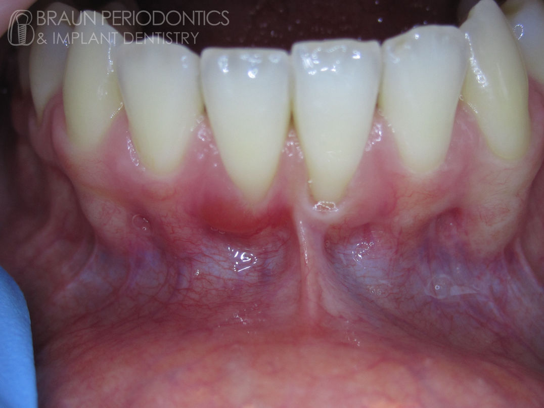
Before: Thing loose tissue on lower teeth
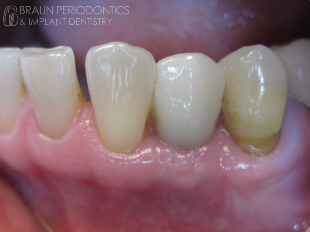
After: Thick border with root coverage
Before: Thin tissue and frenum on lower teeth
After: Thickened tissue with frenum removal
Before: Thin tissue and frenum on lower front tooth
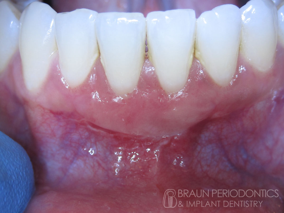
After: Tight healthy non-bleeding gums
Before: Deep pockets and bleeding gums
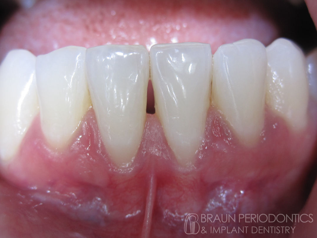
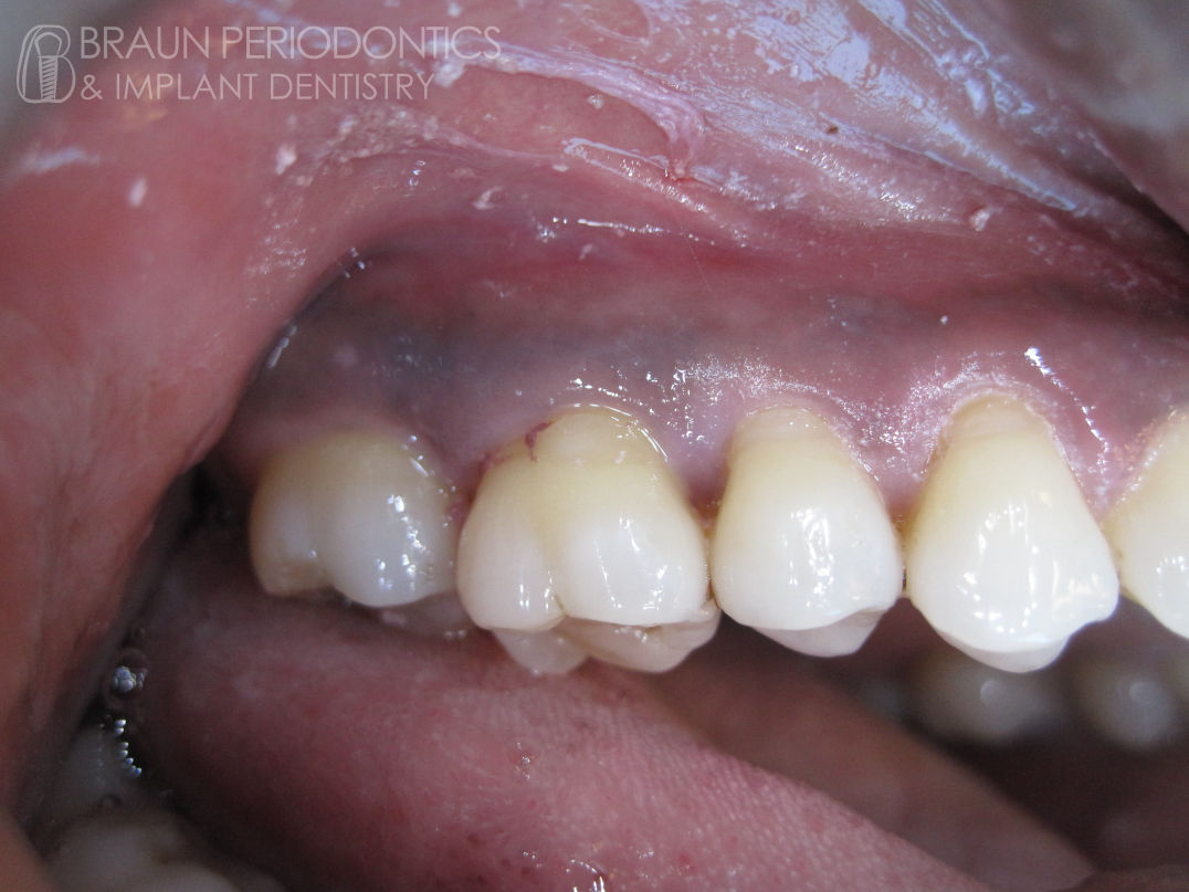
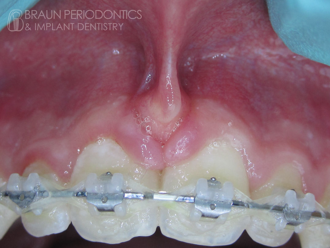
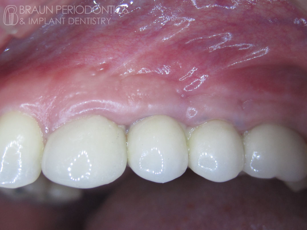
After: Frenum removed and tissues recontoured
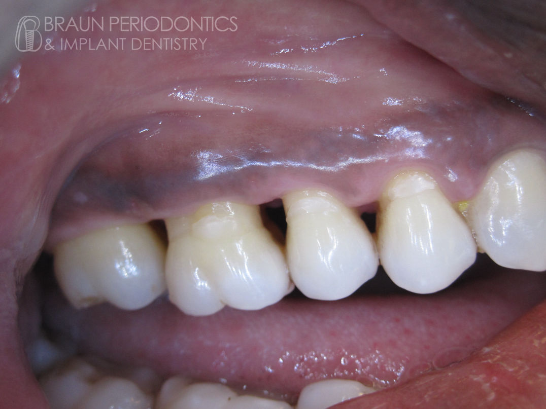
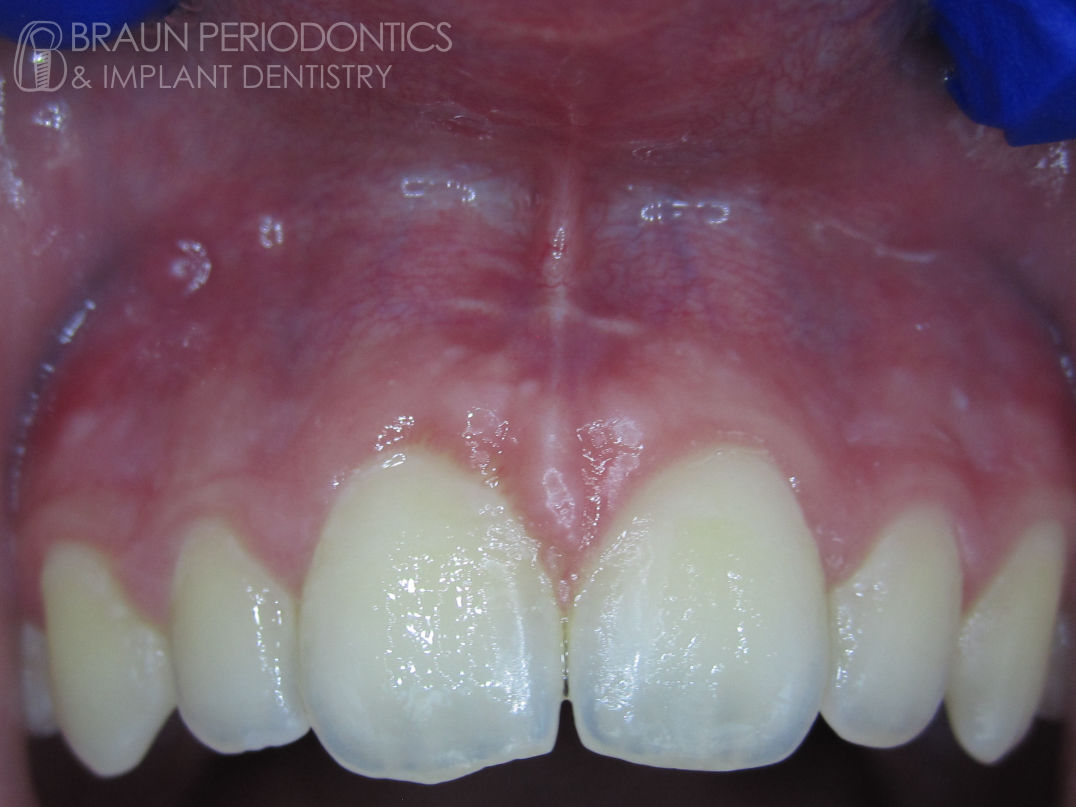
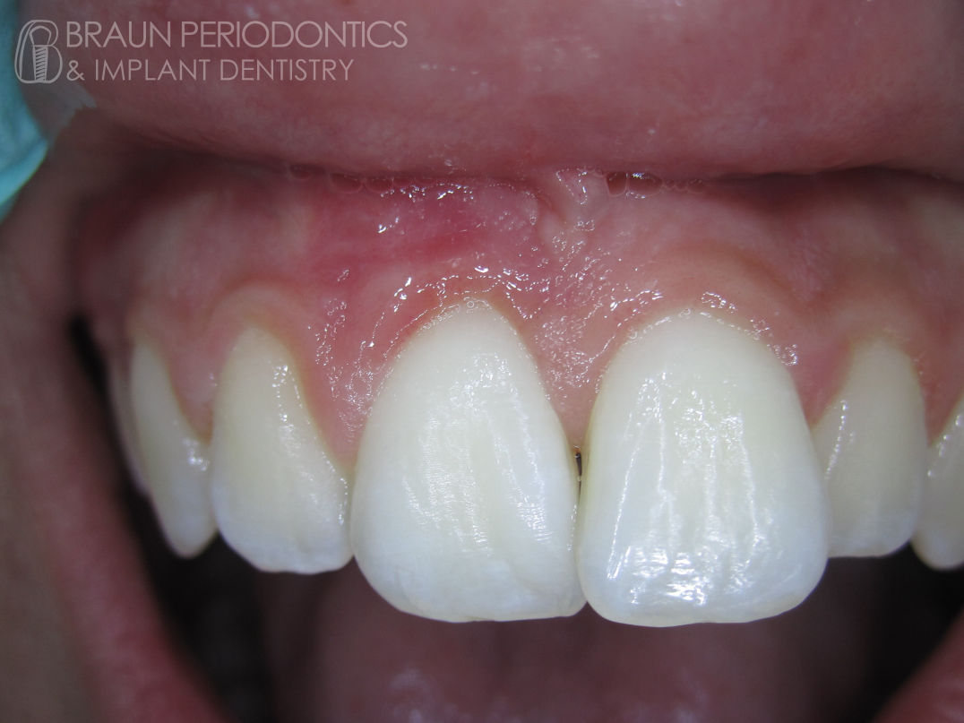
Before: Large prominent upper front frenum
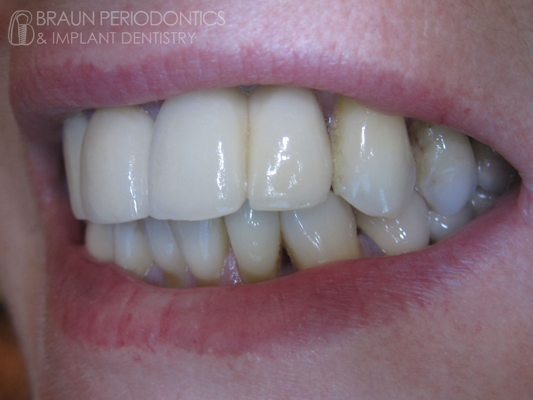
After: Implant bridge to replace 3 teeth
Before: Missing upper back teeth
After: Individual implants replacing 2 teeth
Before: Missing upper front teeth
After: Individual implants replacing 2 teeth
Before: Missing lower back teeth
After: Individual implant replacing tooth
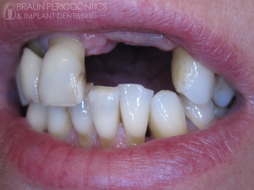
Before: Missing lower canine
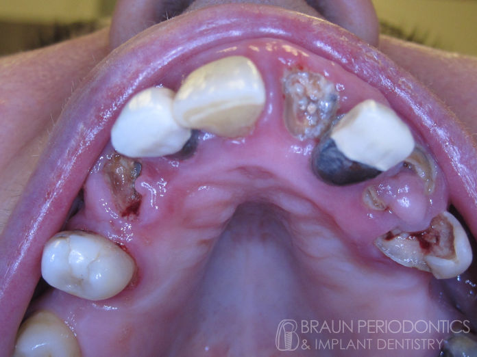
Before: Many decayed teeth, large cavities and roots remaining that were not worth fixing. Most of these teeth were removed and the sockets were grafted with bone to prepare for future implants.
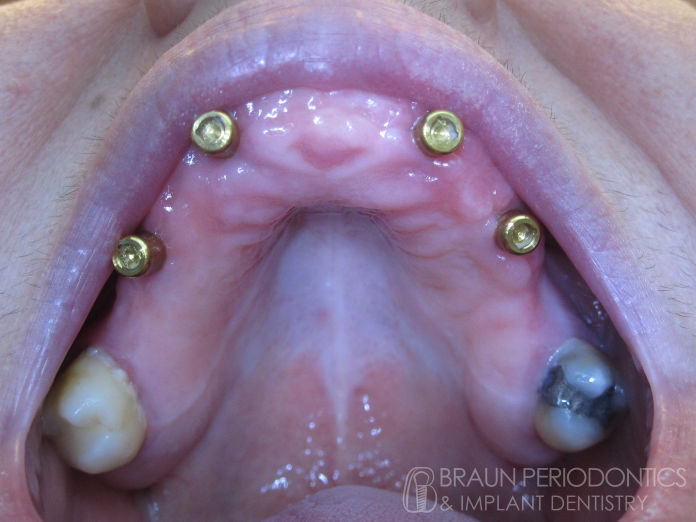
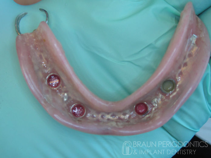
After: 4 implants were placed evenly in the upper jaw, while leaving the 2 molars at the back (which will be removed later if needed). Together, these strong anchors help secure a maxillary denture.
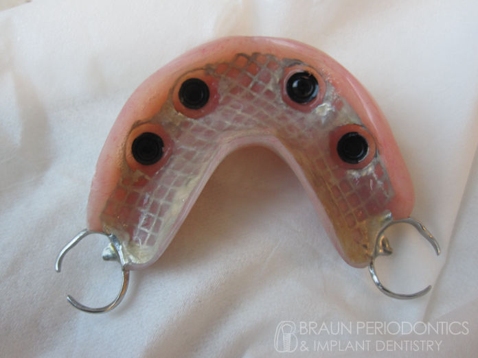
Maxillary denture secured to the 4 implants with no palatal coverage. The clasps at the back grip the 2 remaining molars but they are not needed (eventually these clasps will be removed with the molars).
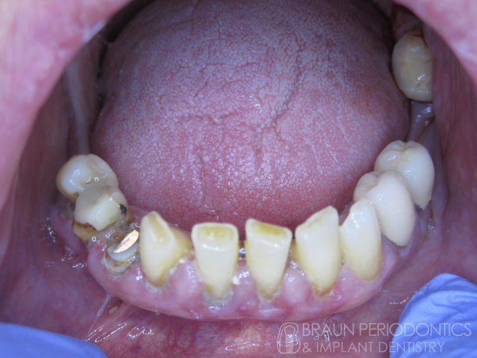
Before: Lower teeth have bone loss, deep pockets, looseness, spacing and cavities, making their prognosis poor. The patient preferred to extract the teeth and place implants.
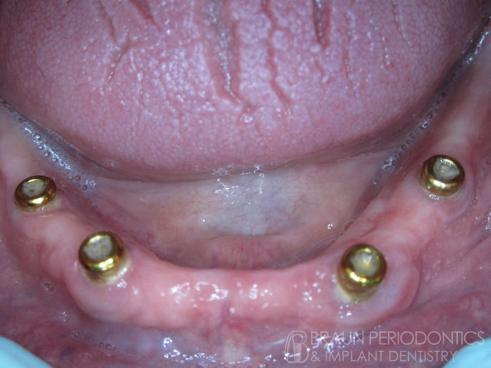
After: Following extractions and bone grafting, 4 implants were placed in the lower arch to provide a stable base for the over-denture. One molar remained (not visible).
The lower denture has 4 attachments which snap on to the gold-colored implant buttons, helping to keep the denture tight and provide a solid foundation.
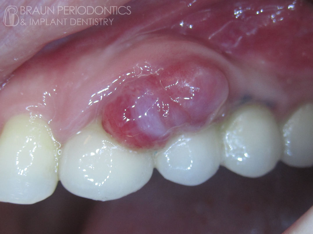
After: Tumor removed and gingiva recontoured
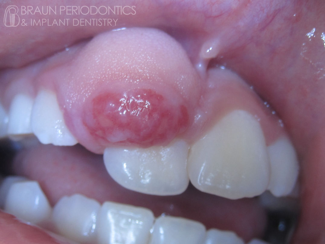
Before: Tumor on the front upper teeth
After: Tumor removed and tissue restored to health
Before: Tumor surrounding a number of implants
Created by Dr. Braun with Mobirise on Ubuntu Studio Linux